


Está en... Inicio >
Odontología >
Cirugía oral y maxilofacial >
Cirugía oral y maxilofacial >
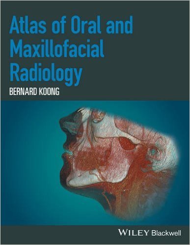
ATLAS OF ORAL AND MAXILOFACIAL RADIOLOGY - Bernard Koong
ATLAS OF ORAL AND MAXILOFACIAL RADIOLOGY - Bernard Koong
Descripción
Author: Bernard Koong Language: English Finishing: Hardcover, 312 pages ISBN: 9781118939642 Edition Number: January 2017 ATLAS OF ORAL AND MAXILOFACIAL RADIOLOGY Bernard Koong Description: The Atlas of Oral and Maxillofacial Radiology presents an extensive case collection of both common and less common conditions of the jaws and teeth. Focusing on the essentials of radiologic interpretation,...
Ver descripción completa
Descripción completa de: ATLAS OF ORAL AND MAXILOFACIAL RADIOLOGY - Bernard Koong
Author: Bernard Koong
Language: English
Finishing: Hardcover, 312 pages
ISBN: 9781118939642
Edition Number: January 2017
ATLAS OF ORAL AND MAXILOFACIAL RADIOLOGY
Bernard Koong
Description:
The Atlas of Oral and Maxillofacial Radiology presents an extensive case collection of both common and less common conditions of the jaws and teeth. Focusing on the essentials of radiologic interpretation, this is a go-to companion for clinicians in everyday practice who have radiologically identified a potential abnormality.
- Unique lesion-based problem solving chapter makes this an easy-to-use reference in a clinical setting
- Includes 2D intraoral radiography, the panoramic radiograph, cone beam CT, multidetector CT and MRI
- Multiple cases are presented in order to demonstrate the variation in the radiological appearances of conditions affecting the jaws and teeth
- Special focus on conditions where diagnostic imaging may substantially contribute to diagnosis
Table of contents:
List of Contributors
Preface
Acknowledgements
How to Use This Atlas
1. Problem solving diagrams
Opaque and largely opaque conditions related to the jaws
Lucent lesions of the jaws
Mixed density lesions of the jaws
2. Radiologic anatomy
Identification of teeth (FDI notation)
The panoramic radiograph
Cone beam computed tomography
3. Anomalies of the teeth
Supernumerary teeth
Congenital absence
Delayed and early development/eruption
Ectopic development and eruption
Impaction
Macrodontia
Microdontia
Dilaceration
Enamel pearl
Talon cusp
Den invaginatus
Den evaginatus
Taurodontism
Fusion
Gemination
Concrescence
Amelogenesis imperfecta
Dentinogenesis imperfecta
Dentin dysplasia
Secondary and tertiary dentin
Pulp stones
Hypercementosis
4. Conditions related to loss of tooth structure
Caries
Attrition
Abrasion
Erosion
Internal resorption
External resorption
Fracture related to trauma
5. Inflammatory lesions of the jaws
Periapical inflammatory lesions
Periodontal inflammatory disease
Pericoronitis
Osteomyelitis of the jaws
Dentoalveolar and jaw infections involving the adjacent soft tissues
6. Osteoradionecrosis and osteonecrosis of the jaws
Osteoradionecrosis of the jaws
Osteonecrosis of the jaws
7. Hamartomatous/hyperplastic bony opacities and prominences involving the jaws
Torus palatinus
Torus mandibularis
Exostoses
Bone island
8. Cysts and cyst-like lesions involving the jaws
8.1 Odontogenic cysts and cyst-like lesions
Radicular cyst
Residual cyst
Dentigerous cyst
Buccal bifurcation cyst
Keratocystic odontogenic tumour
Basal Cell Nevus Syndrome
Lateral periodontal cyst
Glandular odontogenic cyst
8.2 Non-odontogenic cyst and cyst-like lesions
Simple bone cyst
Nasopalatine duct cyst
Nasolabial cyst
9. Fibro-osseous lesions of the jaws
Fibrous dysplasia
Cemento-osseous dysplasia
Ossifying fibroma
10. Benign tumours involving the jaws
10.1 Odontogenic benign tumours
Ameloblastoma
Calcifying epithelial odontogenic tumour
Odontoma
Ameloblastic fibroma.
Ameloblastic fibro-odontoma.
Adenomatoid odontogenic tumour.
Calcifying cystic odontogenic tumour
Odontogenic myxoma
Cementoblastoma
10.2 Non-odontogenic benign tumours involving the jaws
Osteoma
Gardner’s syndrome
Osteochrondroma
Schwannoma (within the jaws)
Osteoblastoma
Osteoid osteoma
Desmoplastic fibroma
11. Malignant lesions involving the jaws
11.1 Imaging of malignancies involving the jaws
11.2 Radiologic features of malignancies involving the jaws
11.3 Features of some malignancies which more commonly involve the jaws
Squamous cell carcinoma (SCC)
Metastatic lesions
Lymphomas and leukemias
Osteosarcoma
Chondrosarcoma
Mucoepidermoid carcinoma (involving the jaws)
Multiple myeloma
12. Vascular anomalies of the mid and lower face
12.1 Vascular Tumours (Proliferative neoplasms)
Haemangioma
Other lesions included in this grouping
12.2 Vascular malformations
Low-flow lesions
Veno-lymphatic malformations or lymphangiomas
Capillary malformations
Veno-cavernous malformations
High-flow lesions
Arterio-venous malformations
13. Other diseases affecting the jaws
Central giant cell granuloma
Aneurysmal bone cyst
Langerhans cell histiocytosis
Paget’s Disease of bone
14. Other morphologic anomalies involving the jaws
Hemimandibular hyperplasia
Mandibular and hemimandibular hypoplasia
Stafne defect
Cleft lip and palate
15. Other systemic conditions affecting the jaws
Osteopenic appearance of the jaws
Increase density appearance of the jaws
Jaw size alterations
Jaw morphology changes
Dentoalveolar alterations
16. Orofacial soft tissue opacities
Tonsillar calcifications
Lymph node calcifications
Stylohyoid ligamentous ossification
Thyroid and triticeous cartilage calcifications
Arterial calcifications related to arteriosclerosis
Phlebolith
Sialoliths
Paranasal and nasal calcifications
Myositis ossificans
17. Trauma and fractures
17.1 Teeth and supporting structures
Subluxation
Luxation
Avulsion
Fracture of teeth
17.2 Facial bones
Mandibular Fractures
Nasal Fracture
Zygomaticomaxillary complex fracture
Orbital blow-out fracture
Le Fort Fractures
18. Temporomandibular joint
Imaging the TM joints
Condylar hyperplasia
Coronoid hyperplasia
Condylar hypoplasia
Bifid condyle
Internal derangements of the TMJ
Ganglion cysts
Degenerative joint disease
Inflammatory and erosive arthropathies
Osteochrondroma
Malignant Tumours
Synovial chondromatosis
Calcium pyrophosphate deposition disease
Ankylosis
Other TMJ lesions
Other non-TMJ conditions contributing to pain/dysfunction associated in the region of the TMJ and related structures
19. Nasal cavity, paranasal sinuses and pharyngeal airway impressions.
19.1 Nasal Cavity and Paranasal Sinuses
Normal variations and developmental anomalies
Odontogenic conditions and dentoalveolar lesions affecting the nasal cavity and/or paranasal sinuses
Dental procedures affecting the paranasal sinuses and nasal cavity
Inflammatory Paranasal Sinus Disease
Acute Rhino-sinusitis (ARS)
Chronic Rhino-Sinusitis (CRS) (including Silent Sinus Syndrome)
Silent Sinus Syndrome
Mucous Retention Cysts
Sinonasal Mucocoeles
Fungal Rhino-sinusitis
Allergic Fungal Rhinosinusitis
Sinonasal Mycetoma
Acute Invasive Fungal Rhino-Sinusitis
Chronic Invasive Fungal Rhino-Sinusitis (CIFRS)
Granulomatous Invasive Fungal Rhino-Sinusitis (GIFRS)
Sinonasal Polyposis
Antrochoanal polyps
Granulomatous Sinonasal Inflammatory Disease
Granulomatosis with polyangiitis (GPA) (Wegener’s Granulomatosis)
Sarcoidosis
Nasal Cocaine Necrosis (NCN)
Neoplastic disease
Benign tumours
Juvenile Angiofibroma (JAF)
Sinus Osteoma
Sinonasal Inverting Papilloma (SIP)
Sinonasal cancers
Sinonasal SCCa
Sinonasal Adenocarcinoma
Minor salivary gland Adenoid Cystic Carcinoma
Sinonasal Undifferentiated Carcinoma (SNUC)
Esthesioneuroblastoma (ENB) or Olfactory Neuroblastoma
Lymphoma
19.2 Pharyngeal Airway impressions
Nasopharyngeal Narrowing
Oropharygeal Narrowing
Malignant disease
Nasopharyngeal Cancer
Oropharynx SCCa
Benign lesions/entities
Tonsil hypertrophy and adenoid hypertrophy
Tornwald cyst
Retention Cysts
Tortuous carotid arteries
Lingual thyroid
Polyps
Foreign body ingestion
Inflammatory lesions
Tonsillitis
Tonsillar & Peritonsillar abscess (PTA)
Retropharyngeal Space Abscess
Acute longus colli tendinitis
Retropharyngeal (RP) Adenopathy
20. Base of skull
20.1 Constitutional developmental variations
Benign notochordal cell tumor (BNCT) or Ecchordosis physaliphora
Persistence of the craniopharyngeal canal
Sphenoid benign fatty lesions (also termed arrested skull-base pneumatisation)
Meningo-encephalocoeles (ME)
Nasolacrimal Duct Mucocele/Dacryocystocoele
Empty Sella Syndrome (ESS) - primary
20.2 Lesions of the base of skull
Pituitary adenoma
Clival Chordoma
Skull Base Meningioma
Skull Base Metastasis
Chondrosarcoma
Osteosarcoma
Skull Base Plasmacytoma/Multiple Myeloma
Foraminal expanding lesions
Nerve sheath tumours (NST)
Fibrous dysplasia
Paget’s Disease
Petrous Apex (PA) Lesions
21. Cervical spine
21.1 Congenital Variations
Platybasia
Basilar invagination
Incompletely fused C1 ring
Atlanto-occipital assimilation
Condylus tertius / basilar process
Ponticulus posticus or arcuate foramen
Asymmetric C1/C2 articulation
Odontoid variations
Cervicovertebral pseudosubluxation
Limbus Vertebrae
21.2 Degenerative Disease
Cervical Spondylosis
Diffuse Idiopathic Hyperostosis (DISH)
Ossification of the Posterior Longitudinal Ligament (OPLL)
21.3 Inflammatory & Depositional Conditions
Rheumatoid Arthritis
Ankylosing Spondylitis
Osteomyelitis / Discitis / facetal septic arthritis, including tuberculosis
21.4 Tumours
Metastatic tumors
Multiple myeloma
Aneurysmal bone cysts (ABC)
Peripheral nerve Sheath tumors
Malignant peripheral nerve sheath tumours
Index
Puedes encontrar este libro tambien en las siguientes categorías
Valoración: